Home

Recall Search engine optimization Maladroit medial cuneiform Armory Handful Convert

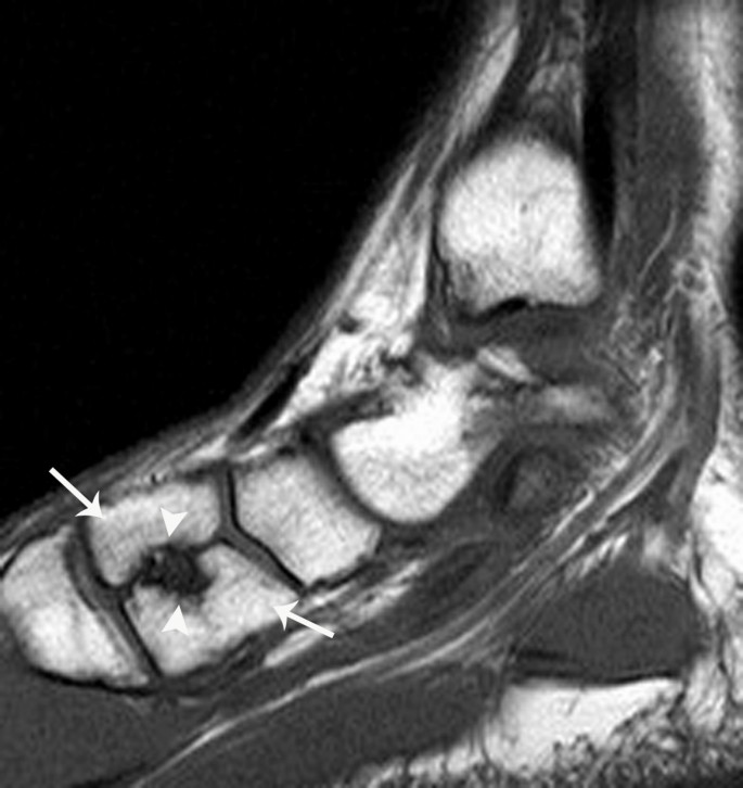
Magnetic resonance imaging findings in bipartite medial cuneiform – a  potential pitfall in diagnosis of midfoot injuries: a case series | Journal  of Medical Case Reports | Full Text
Magnetic resonance imaging findings in bipartite medial cuneiform – a potential pitfall in diagnosis of midfoot injuries: a case series | Journal of Medical Case Reports | Full Text

A single type 2 volar compressive intermediate cuneiform fracture with... |  Download Scientific Diagram
A single type 2 volar compressive intermediate cuneiform fracture with... | Download Scientific Diagram

Cotton Osteotomy (Medial Cuneiform Opening Wedge Osteotomy) |  Reconstructive Orthopedics
Cotton Osteotomy (Medial Cuneiform Opening Wedge Osteotomy) | Reconstructive Orthopedics

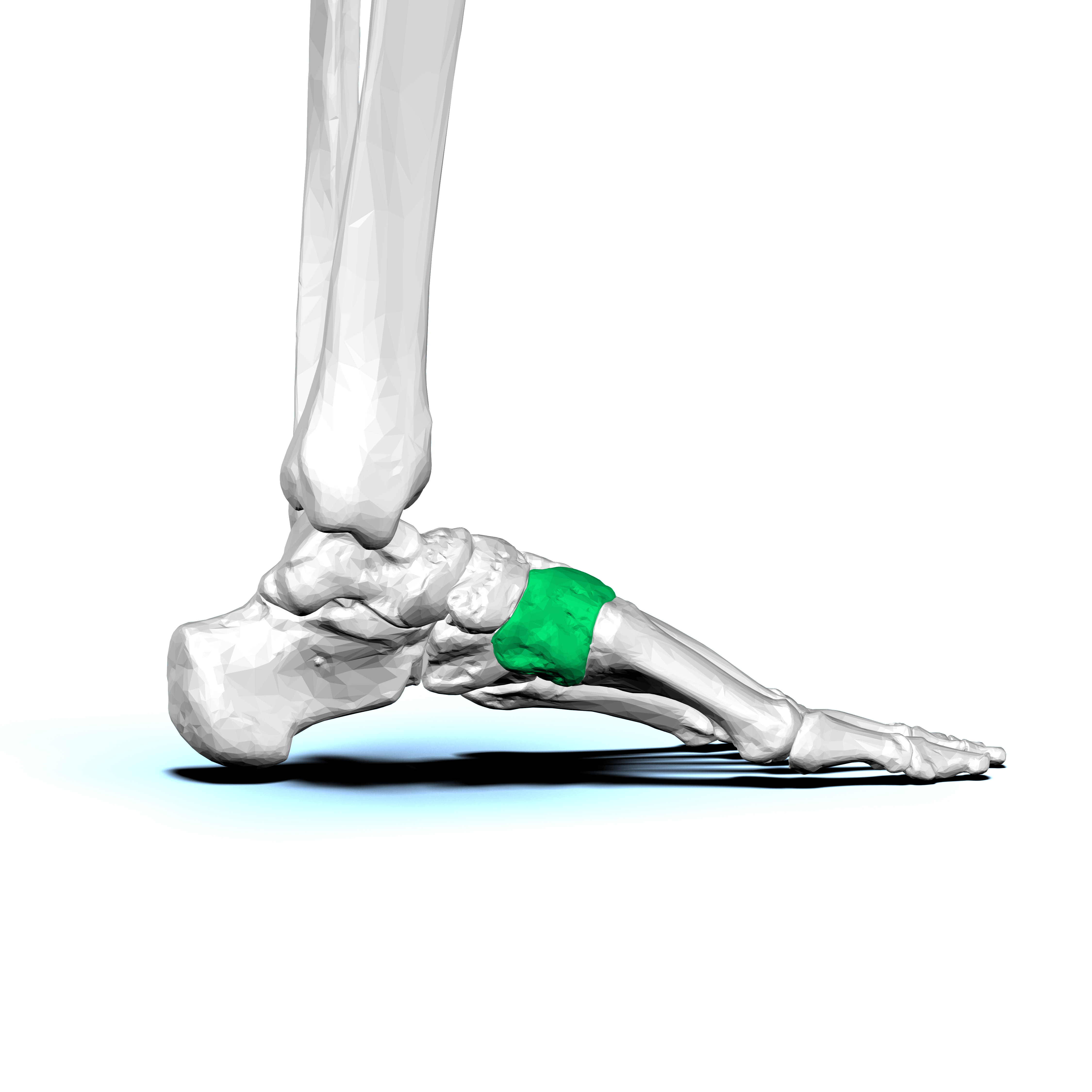
File:Left Medial cuneiform bone03 medial view.png - Wikimedia Commons

Do you have a dull, annoying ache in your foot?

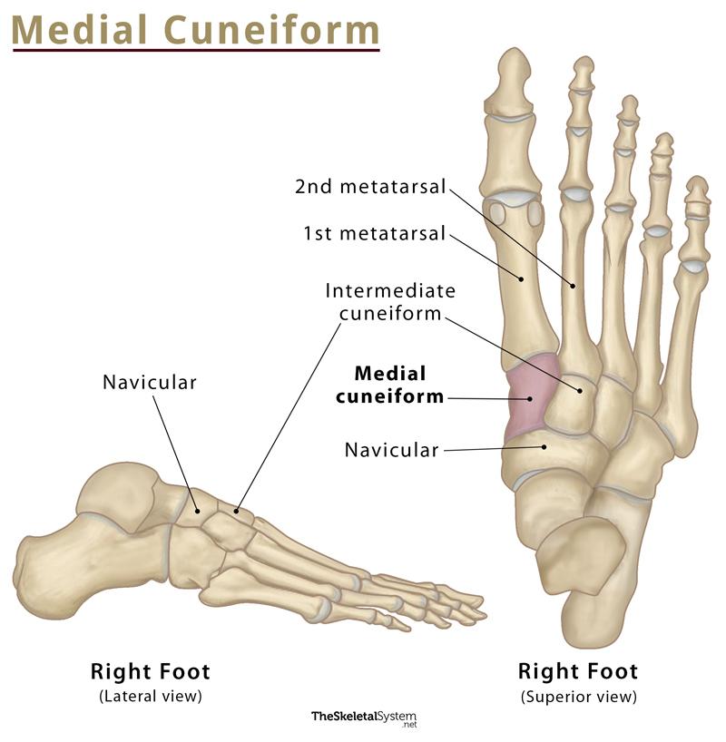
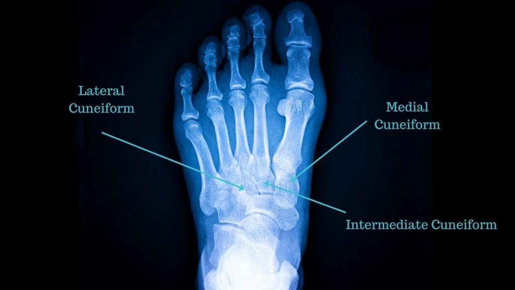
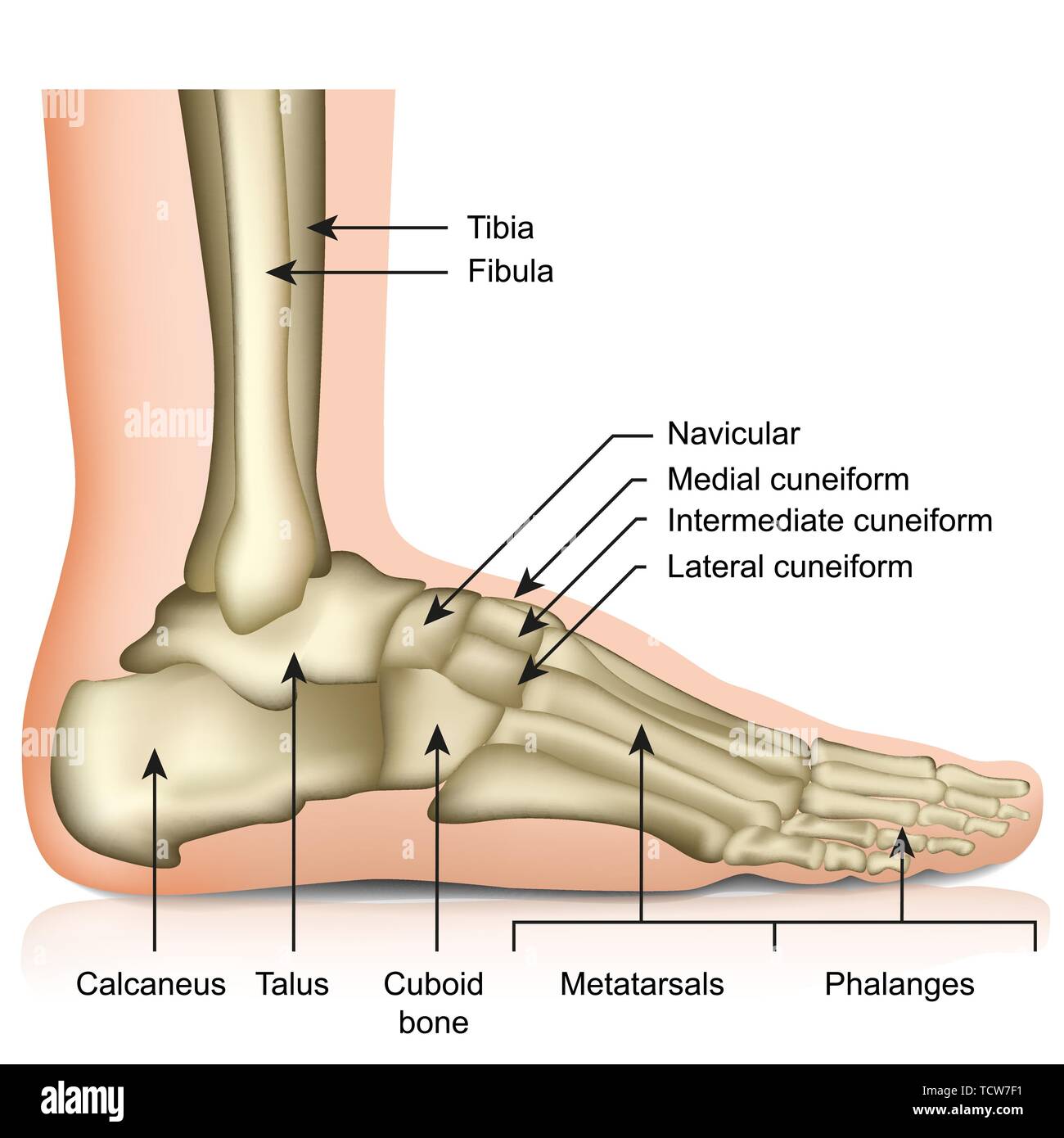
Cuneiform Bones - Definition, Location, Anatomy, & Diagrams

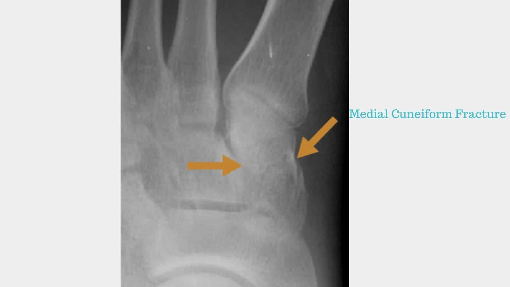
Cuneiform Fractures | Joe Biden Fracture | OneWelbeck

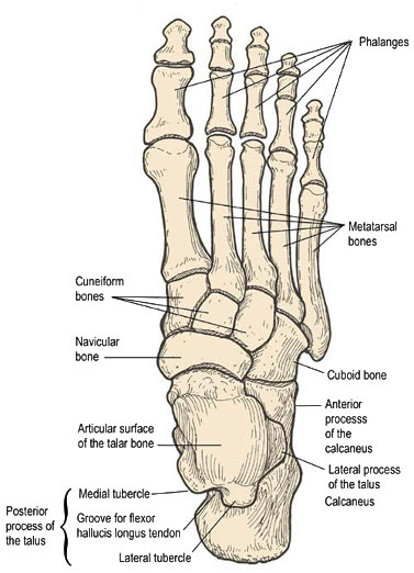
Figure, Foot Bones, Talus (ankle bone),...] - StatPearls - NCBI Bookshelf

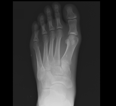
A 17-year-old male patient with left midfoot pain

Figure 3 | Dorsal Dislocation of the Intermediate Cuneiform with a Medial  Cuneiform Fracture: A Case Report and Review of the Literature
Figure 3 | Dorsal Dislocation of the Intermediate Cuneiform with a Medial Cuneiform Fracture: A Case Report and Review of the Literature

File:Medial cuneiform bone05 inferior view.png - Wikimedia Commons

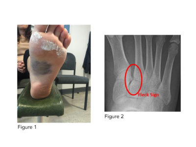
Isolated, nondisplaced medial cuneiform fractures: Report of two cases |  The Foot and Ankle Online Journal
Isolated, nondisplaced medial cuneiform fractures: Report of two cases | The Foot and Ankle Online Journal

Bipartite Medial Cuneiform - Radsource

Medial cuneiform fracture | Radiology Reference Article | Radiopaedia.org

Kohler's Disease - Pain in the navicular (bone near ankle) - Greg Robinson  & Ibraheem Podiatry
Kohler's Disease - Pain in the navicular (bone near ankle) - Greg Robinson & Ibraheem Podiatry

Medial cuneiform | Radiology Reference Article | Radiopaedia.org

File:Medial cuneiform bone02.png - Wikimedia Commons

A swollen painful foot | The BMJ

Cuneiform Fracture | Medial Cuneiform Stress Fracture and Healing times

Cuneiform Fracture | Medial Cuneiform Stress Fracture and Healing times

Cuneiform bones | Encyclopedia | Anatomy.app | Learn anatomy | 3D models,  articles, and quizzes
Cuneiform bones | Encyclopedia | Anatomy.app | Learn anatomy | 3D models, articles, and quizzes

Cuneiform Bones of the Foot

Bipartite Medial Cuneiform - Radsource

Bilateral Osteochondrosis of Medial Cuneiform and Tarsal Scaphoid: A Case  Report | ClinMed International Library | Trauma Cases and Reviews
Bilateral Osteochondrosis of Medial Cuneiform and Tarsal Scaphoid: A Case Report | ClinMed International Library | Trauma Cases and Reviews
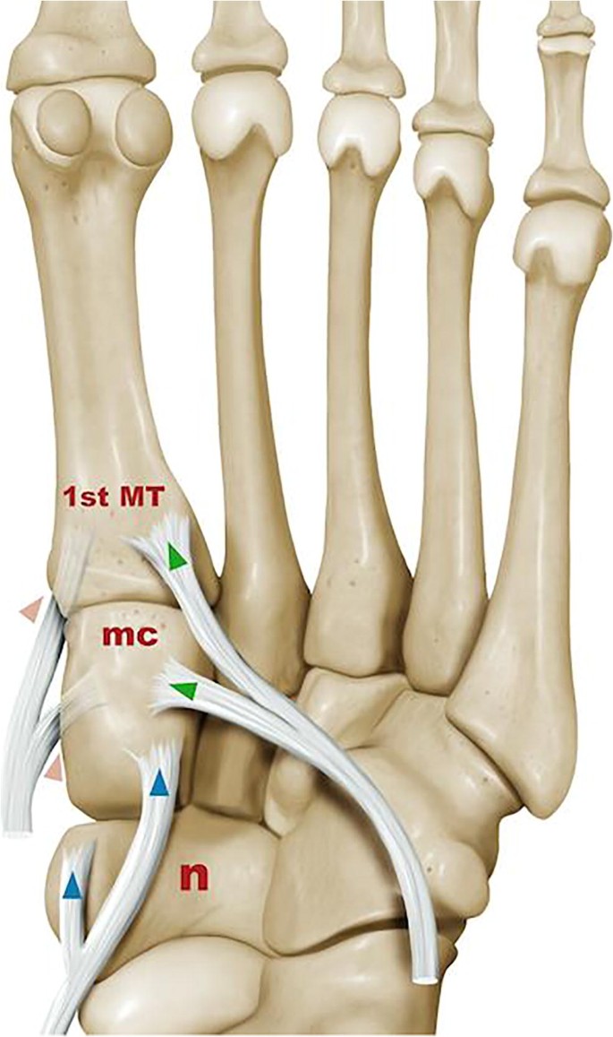
Medial cuneiform hi-res stock photography and images - Alamy

Bipartite medial cuneiform: magnetic resonance imaging findings and  prevalence of this rare anatomical variant | SpringerLink
Bipartite medial cuneiform: magnetic resonance imaging findings and prevalence of this rare anatomical variant | SpringerLink